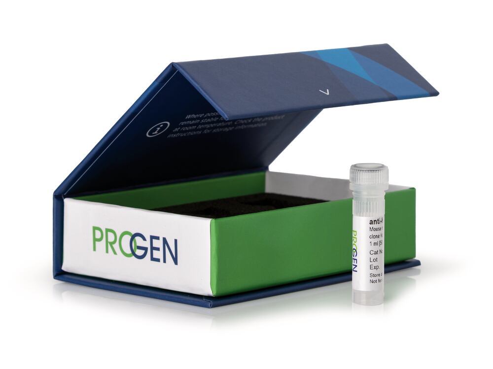
anti-6-His-tag mouse, monoclonal, 6His, after dissolv. in 100 µl PBS, 250 µg/ml, affinity purified (made by PROGEN), Density (D) ~1 g/cm³, Boiling point (bp) 100 °C, Storage temp. +4 °C
Specific primary antibodies that recognise the most commonly used protein tags. The antibodies can be excellently used in cell or tissue biology studies, protein-protein interaction assays, post-translational modifications, expression dynamics or localisation studies, up to the production of analytical kits in laboratory or large scale.
- Available for most commonly used protein tags (His, DDDDK, GST, HA, myc, GFP & Strep)
- High affinity to the respective protein tag
- Robust & highly specific detection of the fusion protein
- Unconjugated
- Suitable for e.g. western blot, immunoprecipitation, ELISA & immunofluorescence
The monoclonal 6-His antibody recognizes polyhistidine (6-His). The 6-His tag is commonly added to recombinant proteins and can be used for detection or purification of the tagged protein.
Application
Immunocytochemistry (ICC): assay dependentImmunoprecipitation (IP): assay dependentWestern Blot (WB): 1:2,000-1:5,000 (0.125-0.05 μg/ml)
Western blot analysis of E. coli lysate containing 6-His-tagged protein with anti-6-His-tag antibody. Western blot analysis was performed on 160 ng, 80 ng or 20 ng of E. coli lysate containing 6-His-tagged protein. Cells were lysed with SDS sample buffer. The PVDF membrane was blocked with 5% dry milk in PBST for 1 h at RT. The primary antibody anti-6-His-tag mouse monoclonal, 6His (Cat. No. 910HISL) was diluted in blocking buffer (antibody concentration 0.125 μg/ml) and incubated for 1 h at RT. The secondary antibody goat anti-mouse IgG polyclonal, HRP conjugate was also diluted in blocking buffer (antibody concentration 0.2 μg/ml) and incubated for 1 h at RT. The bands were visualized by chemiluminescent detection using Pierce™ ECL Western Blotting Substrate.
Western blot analysis of E. coli lysate containing 6-His-tagged protein with anti-6-His-tag antibody. Western blot analysis was performed on 50 ng or 25 ng of E. coli lysate containing 6-His-tagged protein. Cells were lysed with SDS sample buffer. The PVDF membrane was blocked with 5% dry milk in PBST for 1 h at RT. The primary antibody anti-6-His-tag mouse monoclonal, 6His (Cat. No. 910HISL) was diluted in blocking buffer (antibody concentration 0.05 μg/ml) and incubated for 1 h at RT. The secondary antibody goat anti-mouse IgG polyclonal, HRP conjugate was also diluted in blocking buffer (antibody concentration 0.2 μg/ml) and incubated for 1 h at RT. The bands were visualized by chemiluminescent detection using Pierce™ ECL Western Blotting Substrate.
Not a medical device / Not an IVD product
Technical Information
| Application |
ICC/IF IP WB |
| Host species |
mouse |
| Clonality |
monoclonal |
| Purification |
Affinity chromatography |
| Conjugate |
uncojugated |
| Isotype |
IgG1 |
| Clone |
6His |
| Target |
6-His-Tag |