
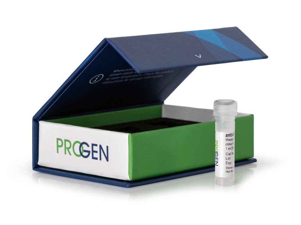

Description
anti-Vimentin mouse monoclonal,, VIM 3B4, 50 µg/ml, affinity purified (made by PROGEN), Density (D) ~1 g/cm³, Boiling point (bp) ~100 °C, Storage temp. +4 °C
This antibody portfolio includes a wide range of tissue- & tumor-specific primary antibodies for basic research, product development and analysis of a variety of biological questions. The monoclonal antibodies are perfectly applicable for all cell or tissue biology studies and for the analysis of predictive markers in research.
- High epitope affinity
- Highly specific detection of the target protein
- Unconjugated
- Suitable for e.g. western blot, immunoprecipitation, ELISA & immunofluorescence
The antibody is highly specific for the intermediate filament protein vimentin which is present in all cells of mesenchymal origin. VIM 3B4 has turned out to be the most avid mab to vimentin. Polypeptide reacting: 57 kDa intermediate filament protein (vimentin) of mesenchymal cells. Tumors specifically detected: sarcoma (including myosarcoma), lymphoma, melanoma. The binding region of monoclonal antibody VIM3B4 has been characterized by Bohn et al.(1992). According to these authors, the epitope has been localized on the alpha-helical part of vimentin (rod domain coil 2). Due to an aa substitution at position of aa 353 in murine vimentin (that could explain for the weak cross-reaction of the antibody with murine vimentin) they were able to narrow down the binding region around position 353. These findings were confirmed by truncation mutagenesis experiments using human vimentin (Rogers et al., 1995). Tested cultured cell lines: fibroblasts (SV-80).
Immunocytochemistry (ICC): assay dependentImmunohistochemistry (IHC) - frozen: 1:100-1:200 (250-500 ng/ml)
| Application | ICC/IF IHC WB |
| Host species | mouse |
| Clonality | monoclonal |
| Reactivity antibody | ape Amphibians |
| Purification | Affinity chromatography |
| Conjugate | uncojugated |
| Concentration | 50 µg/ml |
| Isotype | IgG2a |
| Uniprot ID AK | P48616 (bovine), P09654 (chicken), P08670 (human) |
| Clone | VIM 3B4 |
| Target | Vimentin |