


Description
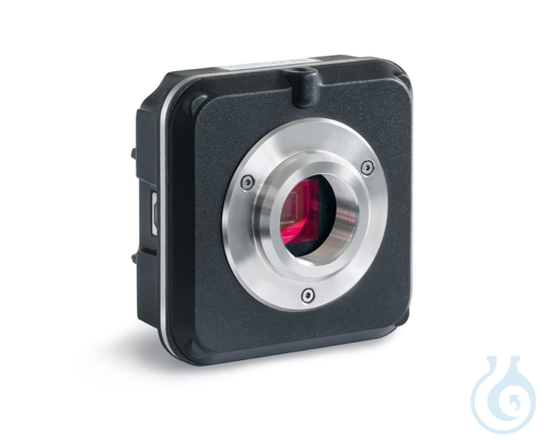
C-Mount camera USB 3.0 ODC 831 is a universal microscope camera intended for measurement, counting, documentation, archiving and image processing. It connects to the microscope and to a laptop or PC via a USB 3.0 cable, with power drawn directly from the USB link so that no external supply is needed. CMOS sensor technology combined with USB 3.0 provides rapid, clear image display and is suitable for advanced methods such as darkfield, phase contrast and fluorescence. Used with the supplied Microscope VIS software, the camera offers strong synchronisation, high frame rate and stable image performance to support efficient daily work. The package from manufacturer Kern includes the camera, multilingual Microscope VIS Basic software KERN OXM 901, a 2000 mm USB cable, various eyepiece adapters and an object micrometer for calibration of the software. Please order the appropriate C-Mount adapter for your microscope at the same time to ensure correct optical coupling.
This article belongs on Microscope objectives, eyepieces & optics, in Microscopy & histology.
Key features
- Pack size: 1 pc(s)
- Interface: USB 3.0
- Sensor technology: CMOS
- USB cable length: 2000 mm
- Software: Microscope VIS Basic OXM 901
- EAN: 4045761190467
- Technical Information Sheet:

Recently viewed
You may also like
Browse related categories
Category
Sub-category
Microscope objectives, eyepieces & optics
Catalogue
Brand
Questions about C-Mount Cameras – USB 3.0 KERN ODC 831
What is this article?
C-Mount Cameras – USB 3.0 KERN ODC 831 is the article on this page. Luminix Health distributes KERN for laboratory use. It sits in Microscope objectives, eyepieces & optics, inside Microscopy & histology, in Lab instruments.
An objective makes the resolution. Magnification and numerical aperture are the two numbers, and the tube length or the infinity correction has to match the microscope. An eyepiece mostly enlarges the image the objective already resolved. A mismatched objective does not become sharper by zooming.
A light microscope forms an image with glass lenses and transmitted or reflected light. A stereo microscope is the lower-magnification instrument for dissecting and for looking at a surface. Fluorescence needs a light source and filters matched to the dye. Histology is the preparation that makes a tissue readable: fixation, a cassette, a section, a stain. The objective's magnification and numerical aperture decide the resolution. The slide and the cover glass decide whether the section sits in the focal plane. Stains and dyes as substances are identified on PubChem. Papers on the methods are on PubMed.
Which category should I open next?
The category is Microscopy & histology. The sub-category is Microscope objectives, eyepieces & optics. The catalogue is Lab instruments.
Shelves beside this one are Microscope slides & cover glasses, Histology cassettes & moulds, Counting chambers & haemocytometers, Stereo microscopes, Light & fluorescence microscopes, Histology reagents & consumables, Microscopy accessories, Cold light sources & lighting and Digital & USB microscopes.
What should I check before I order?
When this page shows a safety data sheet, use that manufacturer file. Hazard text does not belong in the sales description. For a pack size, a grade or a certificate that is not on the page, send a quote request or contact the team.
How does ordering and shipping work?
Luminix Health distributes this article from the heart of Europe. Articles in stock ship within 2–5 business days. Standard delivery is free from €250 excl. VAT. Cold-chain goods and dangerous goods under ADR are never included in that free delivery. The terms are in the Shipping Policy. The full brand index is on shop by brand. Who we are is on about us.
Which other categories should I open?
The seven catalogues are Labware, Lab chemicals, Lab instruments, Lab consumables, Chromatography equipment, Lab safety equipment and Life Science & Microbiology.