
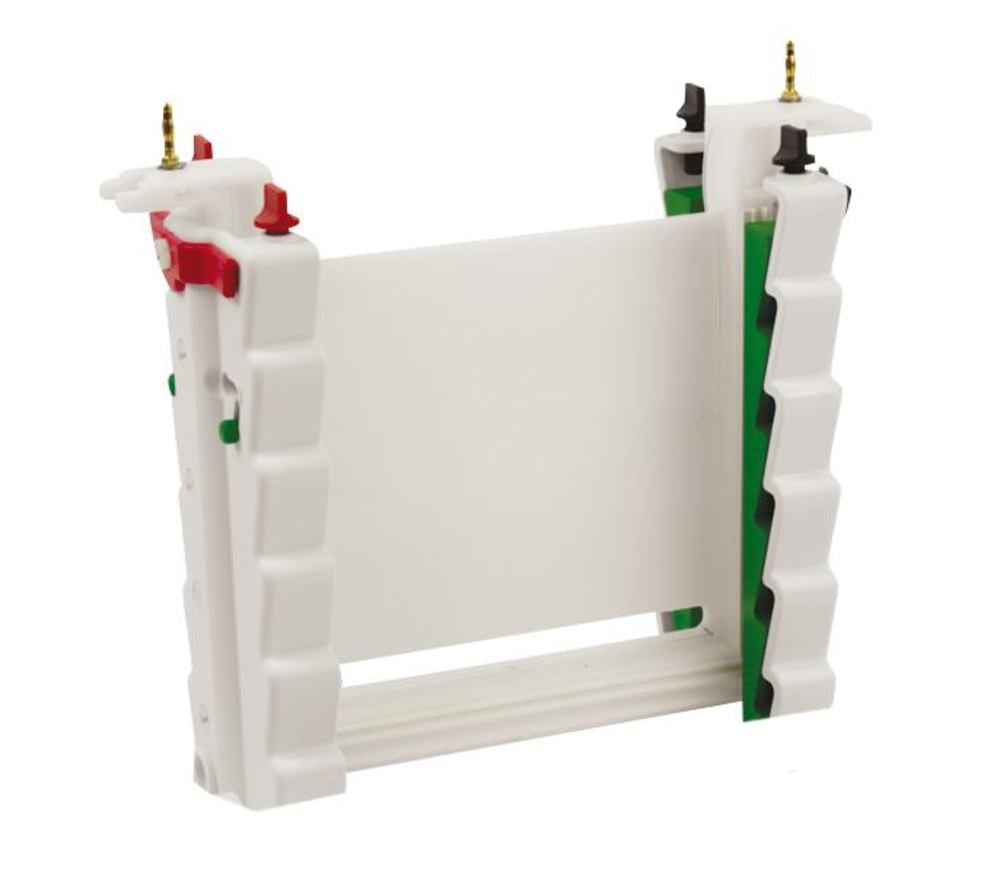


Description
ROTIphorese® PROclamp MAXI is a running module for vertical electrophoresis workflows in the laboratory. Designed as part of the ROTIphorese® PROclamp MAXI system, it supports PAGE and related vertical gel electrophoresis setups where a dedicated running module is required. The unit is supplied as 1 piece and integrates into vertical electrophoresis units and accessories used for protein and nucleic acid separation. Available from manufacturer Carl ROTH, this running module addresses routine gel electrophoresis needs in research, quality control and teaching laboratories that standardise on the PROclamp MAXI format. Clear designation as a running module helps users match the component to existing tanks, clamps and gel cassettes within the same product family. Compact single-unit packaging simplifies ordering and inventory for labs that maintain spare or replacement electrophoresis modules. Application focus remains vertical electrophoresis (PAGE), ensuring the module is selected for the correct separation technique rather than horizontal or capillary systems.
This article is part of Light & fluorescence microscopes, in Microscopy & histology.
Key features
- Pack size: 1 pc
- Product type: Running module
- Brand: ROTIphorese®
- Model: PROclamp MAXI
Recently viewed
You may also like
Questions about ROTIphorese® PROclamp MAXI, Running module, 1 unit(s)
What is this article?
ROTIphorese® PROclamp MAXI, Running module, 1 unit(s) is the article on this page. Luminix Health distributes Carl Roth for laboratory use. It sits in Light & fluorescence microscopes, inside Microscopy & histology, in Lab instruments.
A light microscope looks through a thin specimen. A fluorescence microscope adds a light source and filters matched to a dye, so only the labelled structure shows. The filter set has to match the dye's excitation and emission. A bright-field microscope without those filters cannot do that experiment.
A light microscope forms an image with glass lenses and transmitted or reflected light. A stereo microscope is the lower-magnification instrument for dissecting and for looking at a surface. Fluorescence needs a light source and filters matched to the dye. Histology is the preparation that makes a tissue readable: fixation, a cassette, a section, a stain. The objective's magnification and numerical aperture decide the resolution. The slide and the cover glass decide whether the section sits in the focal plane. Stains and dyes as substances are identified on PubChem. Papers on the methods are on PubMed.
Which category should I open next?
The category is Microscopy & histology. The sub-category is Light & fluorescence microscopes. The catalogue is Lab instruments.
Shelves beside this one are Microscope slides & cover glasses, Microscope objectives, eyepieces & optics, Histology cassettes & moulds, Counting chambers & haemocytometers, Stereo microscopes, Histology reagents & consumables, Microscopy accessories, Cold light sources & lighting and Digital & USB microscopes.
What should I check before I order?
The technical information on this page is the manufacturer's sheet. Read that, not a rewritten specification.
When this page shows a safety data sheet, use that manufacturer file. Hazard text does not belong in the sales description. For a pack size, a grade or a certificate that is not on the page, send a quote request or contact the team.
How does ordering and shipping work?
Luminix Health distributes this article from the heart of Europe. Articles in stock ship within 2–5 business days. Standard delivery is free from €250 excl. VAT. Cold-chain goods and dangerous goods under ADR are never included in that free delivery. The terms are in the Shipping Policy. The full brand index is on shop by brand. Who we are is on about us.
Which other categories should I open?
The seven catalogues are Labware, Lab chemicals, Lab instruments, Lab consumables, Chromatography equipment, Lab safety equipment and Life Science & Microbiology.