


Beschrijving
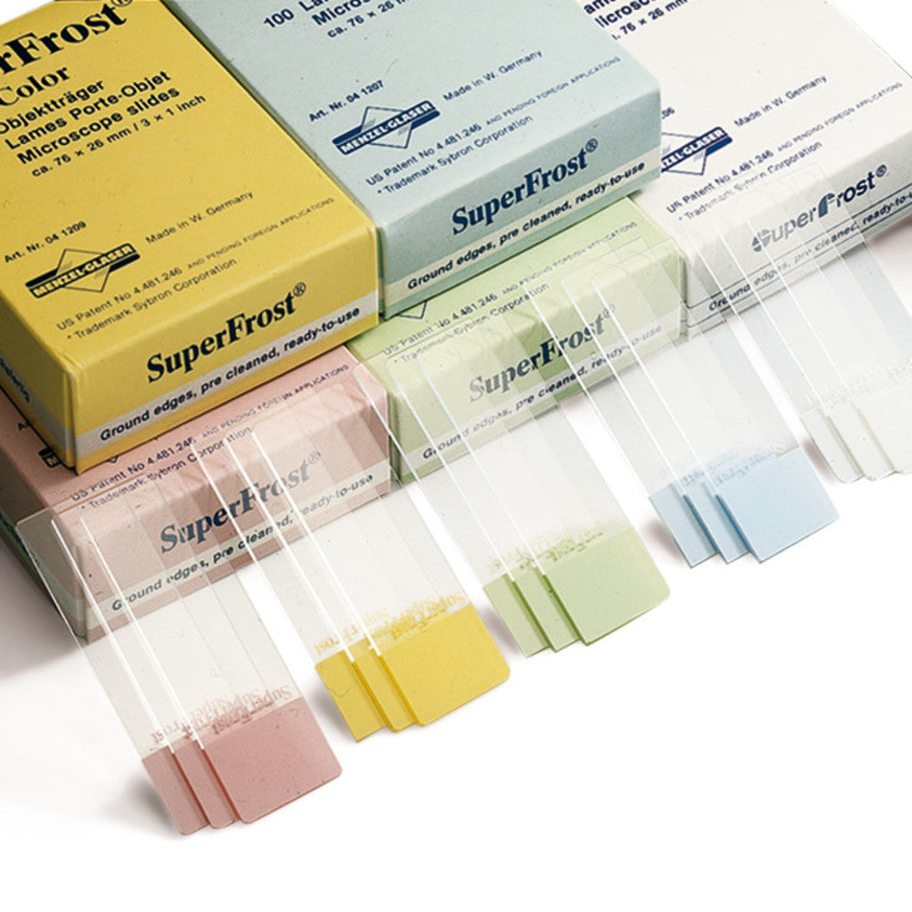
Microscoopglaasjes SuperFrost®, groen zijn standaard laboratoriumglaasjes geproduceerd van natronkalkglas met randen geslepen onder 45°. Het groene SuperFrost®-schrijfvlak biedt een duidelijk kleurgecodeerd oppervlak voor identificatie van specimens in histologie, cytologie en routinemicroscopie. Geslepen randen zorgen voor een veiligere grip en hantering tijdens het monteren, kleuren en afdekken. Elke verpakking bevat 50 stuks en weegt 0,250 kg, een praktisch formaat voor voorraadkasten en gedeelde laboratoriumtrays. Deze glaasjes van fabrikant Epredia zijn bedoeld voor weefselcoupes, uitstrijkjes en vergelijkbare preparaten die een stabiele glazen basis met een duurzaam mat uiteinde voor etikettering vereisen. Groene kleurcodering helpt bij het organiseren van reeksen, parallelle kleuringen en onderwijscollecties. Geschikt voor klinische pathologie, onderzoek en onderwijslaboratoria; de glaasjes sluiten aan bij de standaard lichtmicroscopiepraktijk en gebruikelijke insluitmedia die worden gebruikt in alledaagse histologische en cytologische werkprocessen.
Dit artikel hoort bij Microscoopglaasjes & dekglaasjes, in Microscopie & histologie.
Belangrijkste kenmerken
- Verpakkingsgrootte: 50 stuks
- Producttype: Microscoopglaasjes SuperFrost®
- Kleur: Groen
- Materiaal: Natronkalkglas
- Randen: Geslepen 45°
- Gewicht: 0,250 kg
- Technisch informatieblad:

Recent bekeken
Misschien vind je dit ook leuk
Questions about Microscoopglaasjes SuperFrost®, groen
What is this article?
Microscoopglaasjes SuperFrost®, groen is the article on this page. Luminix Health distributes Epredia for laboratory use. It sits in Microscope slides & cover glasses, inside Microscopy & histology, in Instrumenten.
A slide is the glass a specimen sits on. A cover glass is the thin sheet over it, of a thickness the objective was designed for. A cover glass of the wrong thickness softens the image. Charged and plain slides hold sections differently.
A light microscope forms an image with glass lenses and transmitted or reflected light. A stereo microscope is the lower-magnification instrument for dissecting and for looking at a surface. Fluorescence needs a light source and filters matched to the dye. Histology is the preparation that makes a tissue readable: fixation, a cassette, a section, a stain. The objective's magnification and numerical aperture decide the resolution. The slide and the cover glass decide whether the section sits in the focal plane. Stains and dyes as substances are identified on PubChem. Papers on the methods are on PubMed.
Which category should I open next?
The category is Microscopy & histology. The sub-category is Microscope slides & cover glasses. The catalogue is Instrumenten.
Shelves beside this one are Microscope objectives, eyepieces & optics, Histology cassettes & moulds, Counting chambers & haemocytometers, Stereo microscopes, Light & fluorescence microscopes, Histology reagents & consumables, Microscopy accessories, Cold light sources & lighting and Digital & USB microscopes.
What should I check before I order?
The technical information on this page is the manufacturer's sheet. Read that, not a rewritten specification.
When this page shows a safety data sheet, use that manufacturer file. Hazard text does not belong in the sales description. For a pack size, a grade or a certificate that is not on the page, send a quote request or contact the team.
How does ordering and shipping work?
Luminix Health distributes this article from the heart of Europe. Articles in stock ship within 2–5 business days. Standard delivery is free from €250 excl. VAT. Cold-chain goods and dangerous goods under ADR are never included in that free delivery. The terms are in the Shipping Policy. The full brand index is on shop by brand. Who we are is on about us.
Which other categories should I open?
The seven catalogues are Labware, Lab chemicals, Lab instruments, Lab consumables, Chromatography equipment, Lab safety equipment and Life Science & Microbiology.