
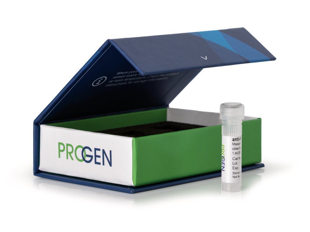

Beschreibung
anti-p53 Protein mouse, monoclonal, Bp53.11, 50 µg/ml, affinity purified (made by PROGEN), Density (D) ~1 g/cm³, Boiling point (bp) ~100 °C, Storage temp. +4 °C
This antibody portfolio includes a wide range of tissue- & tumor-specific primary antibodies for basic research, product development and analysis of a variety of biological questions. The monoclonal antibodies are perfectly applicable for all cell or tissue biology studies and for the analysis of predictive markers in research.
- High epitope affinity
- Highly specific detection of the target protein
- Unconjugated
- Suitable for e.g. western blot, immunoprecipitation, ELISA & immunofluorescence
Excellent marker for wild-type and mutant forms of human p53 antigen. Epitope recognized by Bp53.11: 20-SDLWKLLPENNV-31. The antibody stains positively approx. 60% of investigated carcinoma of lung, breast, colon, stomach, esophagus, pancreas, urinary bladder and testis, head and neck tumors; T-cell Leukemia, non-Hodgkin-Lymphoma, melanoma, sarcoma. Reactivity on cultured cell lines: Hela, MCF-7.
Immunocytochemistry (ICC): assay dependentImmunohistochemistry (IHC) - frozen: 1:1,000-1:3,000 (17-50 ng/ml)Immunohistochemistry (IHC) - paraffin: 1:1,000-1:3,000 (17-50 ng/ml, microwave treatment recommended)Western Blot (WB): 1:2,000-1:10,000 (5-25 ng/ml)
| Application | ICC/IF IHC WB |
| Host species | mouse |
| Clonality | monoclonal |
| Reactivity antibody | human |
| Purification | Affinity chromatography |
| Conjugate | uncojugated |
| Concentration | 50 µg/ml |
| Isotype | IgG2a |
| Uniprot ID AK | Q96A56 (human) |
| Clone | Bp53.11 |
| Target | Recombinant human p53 (transcription domain within the NH2 terminus) |