


Description
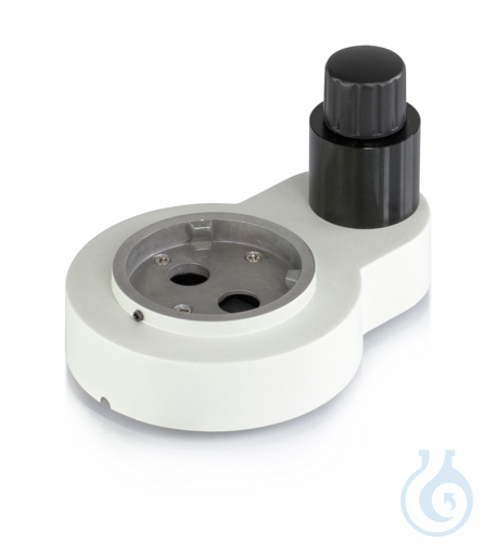
The beam splitter KERN OZB-A5401 is a trinocular beam splitter accessory for stereo microscopy modular systems. It provides a light path division of 100:0, directing the full optical path according to this fixed split ratio for camera or observation routing in trinocular configurations. As a dedicated microscopy accessory, it supports modular stereo microscope setups where a defined beam division is required for documentation, teaching or laboratory imaging workflows. The component is offered as 1 pc(s) and integrates into compatible Kern modular stereo systems without altering the stated 100:0 division characteristic. Net weight is 0,80 kg and gross weight is 1,6 kg, reflecting a compact optical-mechanical unit suitable for bench-top microscope assemblies. This beam splitter from manufacturer Kern belongs to the microscopy accessories category and is intended for laboratories, quality control and educational environments that use trinocular stereo microscope arrangements. Packaging volume and outer dimensions support straightforward storage and handling as a single spare or system expansion part within modular optical setups.
This article is part of Stereo microscopes, in Microscopy & histology.
Key features
- Pack size: 1 pc(s)
- Product type: Trinocular beam splitter
- Beam division: 100:0
- EAN: 4045761169623
- Technical Information Sheet:

Récemment consulté
Vous aimerez peut-être aussi
Browse related categories
Category
Sub-category
Catalogue
Brand
Questions about Beam splitter KERN OZB-A5401
What is this article?
Beam splitter KERN OZB-A5401 is the article on this page. Luminix Health distributes KERN for laboratory use. It sits in Stereo microscopes, inside Microscopy & histology, in Instruments de laboratoire.
A stereo microscope gives a low-magnification, upright, three-dimensional view for dissecting, for sorting and for inspecting a surface. It is not the instrument for a stained section at high power. Working distance and zoom range are the choices.
A light microscope forms an image with glass lenses and transmitted or reflected light. A stereo microscope is the lower-magnification instrument for dissecting and for looking at a surface. Fluorescence needs a light source and filters matched to the dye. Histology is the preparation that makes a tissue readable: fixation, a cassette, a section, a stain. The objective's magnification and numerical aperture decide the resolution. The slide and the cover glass decide whether the section sits in the focal plane. Stains and dyes as substances are identified on PubChem. Papers on the methods are on PubMed.
Which category should I open next?
The category is Microscopy & histology. The sub-category is Stereo microscopes. The catalogue is Instruments de laboratoire.
Shelves beside this one are Microscope slides & cover glasses, Microscope objectives, eyepieces & optics, Histology cassettes & moulds, Counting chambers & haemocytometers, Light & fluorescence microscopes, Histology reagents & consumables, Microscopy accessories, Cold light sources & lighting and Digital & USB microscopes.
What should I check before I order?
When this page shows a safety data sheet, use that manufacturer file. Hazard text does not belong in the sales description. For a pack size, a grade or a certificate that is not on the page, send a quote request or contact the team.
How does ordering and shipping work?
Luminix Health distributes this article from the heart of Europe. Articles in stock ship within 2–5 business days. Standard delivery is free from €250 excl. VAT. Cold-chain goods and dangerous goods under ADR are never included in that free delivery. The terms are in the Shipping Policy. The full brand index is on shop by brand. Who we are is on about us.
Which other categories should I open?
The seven catalogues are Labware, Lab chemicals, Lab instruments, Lab consumables, Chromatography equipment, Lab safety equipment and Life Science & Microbiology.