


Beschrijving
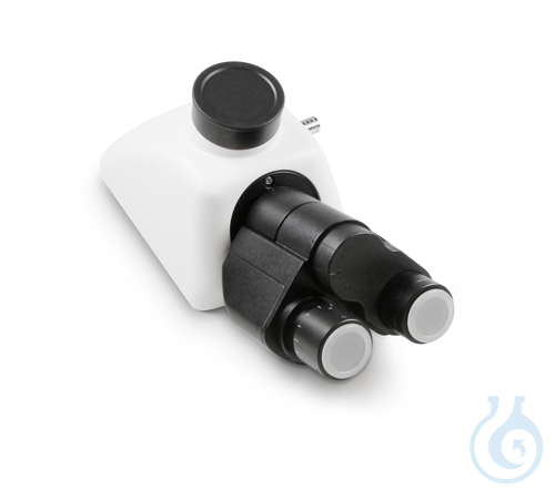
Microscoopkop KERN OBB-A1210 is een trinoculaire tubuskop voor laboratoriummicroscopiesystemen. Ontworpen als een Tubus-Trinokular-samenstel, biedt hij het derde verticale optische pad dat nodig is wanneer binoculair kijken moet worden gecombineerd met camera- of documentatiewerk op een compatibel statief. Dit microscopieaccessoire ondersteunt taken voor kopwisseling of upgrade in onderwijs-, routine- en onderzoekslaboratoria die vertrouwen op een specifieke trinoculaire geometrie. De eenheid wordt geleverd als 1 stuk, met een nettogewicht van 1.4 kg en een brutogewicht van 2 kg. Verpakkingsafmetingen zijn 0.165 m hoogte, 0.265 m breedte en 0.185 m diepte, wat een verpakkingsvolume van 0.008089125 m³ oplevert. De microscoopkop van fabrikant Kern behoort tot de OBB-accessoirefamilie en is bedoeld voor integratie in professionele optische workflows die de bijbehorende KERN-kopinterface accepteren. Als zuiver tubusonderdeel richt hij zich op het observatie- en beeldvormingspad zonder verlichtings- of objecttafelfuncties toe te voegen. Laboratoria die trinoculaire koppen specificeren voor gestructureerde beeldvorming kunnen de OBB-A1210 gebruiken als een gedefinieerd vervangings- of uitbreidingsonderdeel binnen het KERN-microscopieassortiment.
Dit artikel maakt deel uit van Licht- & fluorescentiemicroscopen, in Microscopie & histologie.
Kenmerken
- Verpakkingsgrootte: 1 stuk
- Producttype: Trinoculaire microscoopkop
- Configuratie: Trinoculaire tubus (Tubus-Trinokular)
- Serie: KERN OBB
- EAN: 4045761167421
- Technisch datablad:

Recent bekeken
Misschien vind je dit ook leuk
Questions about Microscoopkop KERN OBB-A1210
What is this article?
Microscoopkop KERN OBB-A1210 is the article on this page. Luminix Health distributes Kern for laboratory use. It sits in Licht- & fluorescentiemicroscopen, inside Microscopie & histologie, in Instrumenten.
A light microscope looks through a thin specimen. A fluorescence microscope adds a light source and filters matched to a dye, so only the labelled structure shows. The filter set has to match the dye's excitation and emission. A bright-field microscope without those filters cannot do that experiment.
A light microscope forms an image with glass lenses and transmitted or reflected light. A stereo microscope is the lower-magnification instrument for dissecting and for looking at a surface. Fluorescence needs a light source and filters matched to the dye. Histology is the preparation that makes a tissue readable: fixation, a cassette, a section, a stain. The objective's magnification and numerical aperture decide the resolution. The slide and the cover glass decide whether the section sits in the focal plane. Stains and dyes as substances are identified on PubChem. Papers on the methods are on PubMed.
Which category should I open next?
The category is Microscopie & histologie. The sub-category is Licht- & fluorescentiemicroscopen. The catalogue is Instrumenten.
Shelves beside this one are Objectglaasjes & dekglaasjes, Microscoopobjectieven, oculairs & optiek, Histologiecassettes & mallen, Telkamers & hemocytometers, Stereomicroscopen, Histologiereagentia & verbruiksartikelen, Microscopieaccessoires, Koudlichtbronnen & verlichting and Digitale & USB-microscopen.
What should I check before I order?
When this page shows a safety data sheet, use that manufacturer file. Hazard text does not belong in the sales description. For a pack size, a grade or a certificate that is not on the page, send a quote request or contact the team.
How does ordering and shipping work?
Luminix Health distributes this article from the heart of Europe. Articles in stock ship within 2–5 business days. Standard delivery is free from €250 excl. VAT. Cold-chain goods and dangerous goods under ADR are never included in that free delivery. The terms are in the Shipping Policy. The full brand index is on shop by brand. Who we are is on about us.
Which other categories should I open?
The seven catalogues are Labware, Lab chemicals, Lab instruments, Lab consumables, Chromatography equipment, Lab safety equipment and Life Science & Microbiology.