


Description
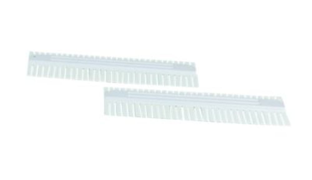
ROTIPHORESE® PROclamp MAXI Comb is a comb accessory for vertical electrophoresis (PAGE), specified with a thickness of 2,0 mm, 5 wells and a well volume of 400 µl. This layout balances moderate sample throughput with a relatively large loading volume on MAXI-format polyacrylamide gels, supporting both analytical comparison and higher-load applications. Five clearly defined wells help maintain consistent lane spacing and gel geometry during casting, which is important for reproducible band patterns after separation. Supplied as 1 pc, the comb is designed for PROclamp MAXI vertical electrophoresis units and compatible accessories used in protein and nucleic acid laboratories. From manufacturer ROTH SELECTION, the 2,0 mm thickness matches thicker gel setups where mechanical stability and increased well capacity are preferred. Select this 5-well, 400 µl comb when you need fewer lanes than high-density formats but greater volume per well for MAXI PAGE workflows.
This article is part of Light & fluorescence microscopes, in Microscopy & histology.
Key features
- Pack size: 1 pc
- Product type: PROclamp MAXI electrophoresis comb
- Thickness: 2,0 mm
- Number of wells: 5
- Well volume: 400 µl
- Format: MAXI vertical electrophoresis
Recently viewed
You may also like
Questions about ROTIPHORESE® PROclamp MAXI Comb, thickness 2.0 mm, 5 wells, 400 µl, 1 unit(s)
What is this article?
ROTIPHORESE® PROclamp MAXI Comb, thickness 2.0 mm, 5 wells, 400 µl, 1 unit(s) is the article on this page. Luminix Health distributes Carl Roth for laboratory use. It sits in Light & fluorescence microscopes, inside Microscopy & histology, in Lab instruments.
A light microscope looks through a thin specimen. A fluorescence microscope adds a light source and filters matched to a dye, so only the labelled structure shows. The filter set has to match the dye's excitation and emission. A bright-field microscope without those filters cannot do that experiment.
A light microscope forms an image with glass lenses and transmitted or reflected light. A stereo microscope is the lower-magnification instrument for dissecting and for looking at a surface. Fluorescence needs a light source and filters matched to the dye. Histology is the preparation that makes a tissue readable: fixation, a cassette, a section, a stain. The objective's magnification and numerical aperture decide the resolution. The slide and the cover glass decide whether the section sits in the focal plane. Stains and dyes as substances are identified on PubChem. Papers on the methods are on PubMed.
Which category should I open next?
The category is Microscopy & histology. The sub-category is Light & fluorescence microscopes. The catalogue is Lab instruments.
Shelves beside this one are Microscope slides & cover glasses, Microscope objectives, eyepieces & optics, Histology cassettes & moulds, Counting chambers & haemocytometers, Stereo microscopes, Histology reagents & consumables, Microscopy accessories, Cold light sources & lighting and Digital & USB microscopes.
What should I check before I order?
The technical information on this page is the manufacturer's sheet. Read that, not a rewritten specification.
When this page shows a safety data sheet, use that manufacturer file. Hazard text does not belong in the sales description. For a pack size, a grade or a certificate that is not on the page, send a quote request or contact the team.
How does ordering and shipping work?
Luminix Health distributes this article from the heart of Europe. Articles in stock ship within 2–5 business days. Standard delivery is free from €250 excl. VAT. Cold-chain goods and dangerous goods under ADR are never included in that free delivery. The terms are in the Shipping Policy. The full brand index is on shop by brand. Who we are is on about us.
Which other categories should I open?
The seven catalogues are Labware, Lab chemicals, Lab instruments, Lab consumables, Chromatography equipment, Lab safety equipment and Life Science & Microbiology.