


Description
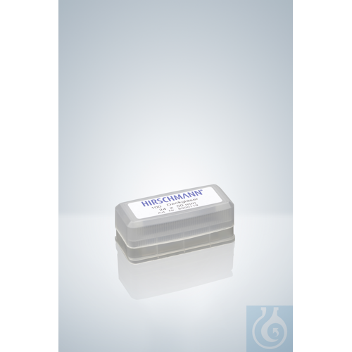
Cover glasses for microscopy 22 x 50 mm with a thickness of 0,13 - 0,17 mm are precision coverslips intended for microscopic specimen preparation and examination. Manufactured to DIN 58 884 and CE marked according to IVD 98/79 EG, they fulfil established specifications for in vitro diagnostic microscopy applications. The 22 mm by 50 mm rectangular format accommodates longer specimen areas on standard microscope slides, and the tightly specified thickness of 0,13 - 0,17 mm supports reliable optical performance during observation. Available in packs of 1000 pieces from manufacturer Hirschmann, these cover glasses suit high-throughput workflows in histology, cytology and general laboratory microscopy. They form a clear protective layer over wet mounts and prepared sections, helping to hold samples in place under the microscope. With a net weight of 0,448 kg per pack and origin in Germany, the product offers a consistent consumable for clinical, research and teaching laboratories that depend on uniform cover-glass size and thickness for routine imaging.
This article is part of Microscope slides & cover glasses, in Microscopy & histology.
Key features
- Pack size: 1000 pc(s)
- Storage temperature: +5 - +40 °C
- Dimensions: 22 x 50 mm
- Thickness: 0,13 - 0,17 mm
- Standard: DIN 58 884
- CE marking: IVD 98/79 EG
Recently viewed
You may also like
Questions about COVER GLASSES FOR MICROSCOPY, 22 X 50 MM, T 0,13 - 0,17 MM
What is this article?
COVER GLASSES FOR MICROSCOPY, 22 X 50 MM, T 0,13 - 0,17 MM is the article on this page. Luminix Health distributes Hirschmann for laboratory use. It sits in Microscope slides & cover glasses, inside Microscopy & histology, in Lab instruments.
A slide is the glass a specimen sits on. A cover glass is the thin sheet over it, of a thickness the objective was designed for. A cover glass of the wrong thickness softens the image. Charged and plain slides hold sections differently.
A light microscope forms an image with glass lenses and transmitted or reflected light. A stereo microscope is the lower-magnification instrument for dissecting and for looking at a surface. Fluorescence needs a light source and filters matched to the dye. Histology is the preparation that makes a tissue readable: fixation, a cassette, a section, a stain. The objective's magnification and numerical aperture decide the resolution. The slide and the cover glass decide whether the section sits in the focal plane. Stains and dyes as substances are identified on PubChem. Papers on the methods are on PubMed.
Which category should I open next?
The category is Microscopy & histology. The sub-category is Microscope slides & cover glasses. The catalogue is Lab instruments.
Shelves beside this one are Microscope objectives, eyepieces & optics, Histology cassettes & moulds, Counting chambers & haemocytometers, Stereo microscopes, Light & fluorescence microscopes, Histology reagents & consumables, Microscopy accessories, Cold light sources & lighting and Digital & USB microscopes.
What should I check before I order?
When this page shows a safety data sheet, use that manufacturer file. Hazard text does not belong in the sales description. For a pack size, a grade or a certificate that is not on the page, send a quote request or contact the team.
How does ordering and shipping work?
Luminix Health distributes this article from the heart of Europe. Articles in stock ship within 2–5 business days. Standard delivery is free from €250 excl. VAT. Cold-chain goods and dangerous goods under ADR are never included in that free delivery. The terms are in the Shipping Policy. The full brand index is on shop by brand. Who we are is on about us.
Which other categories should I open?
The seven catalogues are Labware, Lab chemicals, Lab instruments, Lab consumables, Chromatography equipment, Lab safety equipment and Life Science & Microbiology.